Transverse cervical nerve
| Transverse cervical nerve | |
|---|---|
 The nerves of the scalp, face, and side of neck. ("Cervical cutaneous" identified at center.) | |
 Plan of the cervical plexus. ("Superficial cervical" labeled at center.) | |
| Details | |
| From | Cervical plexus (C2 and C3) |
| Innervates | Cutaneous innervation of the anterior and lateral parts of the neck |
| Identifiers | |
| Latin | nervus transversus colli |
| TA98 | Lua error in Module:Wikidata at line 746: attempt to index field 'wikibase' (a nil value). |
| TH | {{#property:P1694}} |
| TE | {{#property:P1693}} |
| FMA | {{#property:P1402}} |
| Anatomical terms of neuroanatomy [[[d:Lua error in Module:Wikidata at line 865: attempt to index field 'wikibase' (a nil value).|edit on Wikidata]]] | |
The transverse cervical nerve (superficial cervical or cutaneous cervical) is a cutaneous (sensory) nerve of the cervical plexus that arises from the second and third cervical spinal nerves (C2-C3). It curves around the posterior border of the sternocleidomastoideus muscle, then pierces the fascia of the neck before dividing into two branches. It provides sensory innervation to the front of the neck.[1]
Anatomy
[edit | edit source]Course and relations
[edit | edit source]It curves around the posterior border of the sternocleidomastoideus muscle[1] about its middle, and, passing obliquely forward beneath the external jugular vein to the anterior border of the muscle,[citation needed] it perforates the deep cervical fascia before dividing into an ascending branch and a descending branch[1] beneath the platysma.[citation needed] The ascending branch communicates with the cervical branch of the facial nerve.[1]
Dissection
[edit | edit source]During dissection, the sternocleidomastoid muscle is the landmark, with the transverse cervical nerve passing horizontally over this muscle from Erb's point.[citation needed]
Distribution
[edit | edit source]The nerve provides sensory innervation to the skin of the anterior neck between the chin and the sternum.[1]
Additional images
[edit | edit source]-
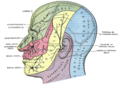 Dermatome distribution of the trigeminal nerve
 -
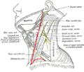 Side of neck, showing chief surface markings.


References
[edit | edit source] This article incorporates text in the public domain from the 20th edition of Gray's Anatomy (1918)
External links
[edit | edit source]- Anatomy figure: 25:03-07 at Human Anatomy Online, SUNY Downstate Medical Center
- lesson6 at The Anatomy Lesson by Wesley Norman (Georgetown University)
Lua error in mw.title.lua at line 392: bad argument #2 to 'title.new' (unrecognized namespace name 'Portal'). Lua error in Module:Authority_control at line 153: attempt to index field 'wikibase' (a nil value).
 | This neuroanatomy article is a stub. You can help Wikipedia by expanding it. |