Popliteal fossa
| Popliteal fossa | |
|---|---|
 Popliteal fossa of the right leg. | |
| Details | |
| Identifiers | |
| Latin | fossa poplitea |
| TA98 | Lua error in Module:Wikidata at line 746: attempt to index field 'wikibase' (a nil value). |
| TH | {{#property:P1694}} |
| TE | {{#property:P1693}} |
| FMA | {{#property:P1402}} |
| Anatomical terminology [[[d:Lua error in Module:Wikidata at line 865: attempt to index field 'wikibase' (a nil value).|edit on Wikidata]]] | |
The popliteal fossa (also referred to as hough or kneepit in analogy to the cubital fossa) is a shallow depression located at the back of the knee joint. The bones of the popliteal fossa are the femur and the tibia. Like other flexion surfaces of large joints (groin, armpit, cubital fossa and essentially the anterior part of the neck), it is an area where blood vessels and nerves pass relatively superficially, and with an increased number of lymph nodes.
Structure
[edit | edit source]Boundaries
[edit | edit source]The boundaries of the fossa are:[1]
| Medial | Lateral | |
|---|---|---|
| Superior | the semimembranosus & semitendinosus muscles[2] | the biceps femoris muscle[2] |
| Inferior | the medial head of the gastrocnemius muscle[2] | the lateral head of the gastrocnemius muscle and plantaris muscle[2] |
Roof
[edit | edit source]Moving from superficial to deep structures, the roof is formed by:
- the skin.[1]
- the superficial fascia.[1] This contains the small saphenous vein, the terminal branch of the posterior cutaneous nerve of the thigh, posterior division of the medial cutaneous nerve, lateral sural cutaneous nerve, and medial sural cutaneous nerve.[1]
- the popliteal fascia.[1][2]
Floor
[edit | edit source]The floor is formed by:
- the popliteal surface of the femur.[2]
- the capsule of the knee joint and the oblique popliteal ligament.[2]
- strong fascia covering the popliteus muscle.[2]
Contents
[edit | edit source]Structures within the popliteal fossa include, (from superficial to deep):[1]
- tibial nerve
- common fibular nerve (also known as the common peroneal nerve)[3]
- popliteal vein
- popliteal artery, a continuation of the femoral artery
- small saphenous vein (termination)[3]
- Popliteal lymph nodes and vessels[3]
It is of note that the common fibular nerve also begins at the superior angle of the popliteal fossa.[4]
Additional images
[edit | edit source]-
 Muscles of the gluteal and posterior femoral regions.
 -
 Small saphenous vein and its tributaries.
 -
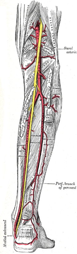 The popliteal, posterior tibial, and peroneal arteries.
 -
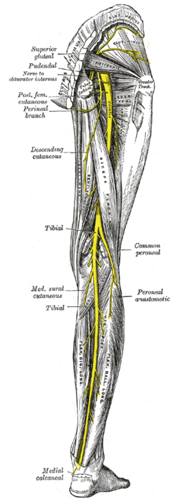 Nerves of the right lower extremity. Posterior view.
 -
 Muscles of thigh. Lateral view.


See also
[edit | edit source]References
[edit | edit source]- ^ a b c d e f Lua error in Module:Citation/CS1/Configuration at line 2172: attempt to index field '?' (a nil value).
- ^ a b c d e f g h Lua error in Module:Citation/CS1/Configuration at line 2172: attempt to index field '?' (a nil value).
- ^ a b c Clinically Oriented Anatomy by Moore, 6th edition
- ^ Lua error in Module:Citation/CS1/Configuration at line 2172: attempt to index field '?' (a nil value).
External links
[edit | edit source]
- postthigh at The Anatomy Lesson by Wesley Norman (Georgetown University) (poplitealfossabones, poplitealfossacontents, poplitealfossafloor)
- MedicalMnemonics.com: 2747 9


Lua error in Module:Authority_control at line 153: attempt to index field 'wikibase' (a nil value).